MR Physics

Overview

The MR Physics group is working on the development of new and improved methods for magnetic resonance imaging (MRI) and spectroscopy (MRS) that aim to provide a non-invasive access to the structure, organization, function, and metabolism of the human central nervous system in vivo.
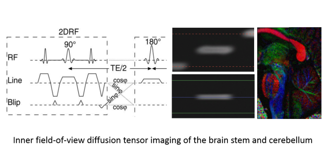
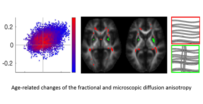
A major focus of our work is diffusion-weighted MRI, the localized measurement of water diffusion properties in tissue that are sensitive to the tissue structure on a cellular or microscopic scale. With extended diffusion-weighting methods like the double diffusion encoding used in our group, more specific information like the density, size, and shape of the cells may become accessible and could help to distinguish and characterize healthy and pathological tissue. Furthermore, we aim to speed-up and improve the image quality of diffusion-weighted MRI, in particular for applications in the spinal cord to make it clinically feasible for patients with spinal cord lesions or injury.

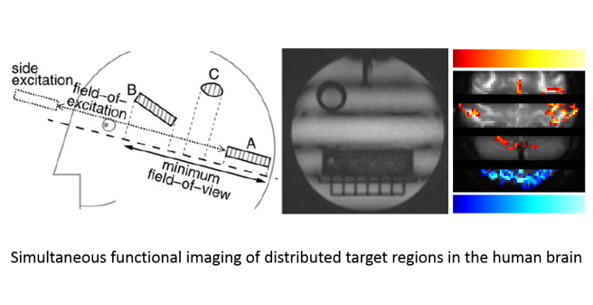
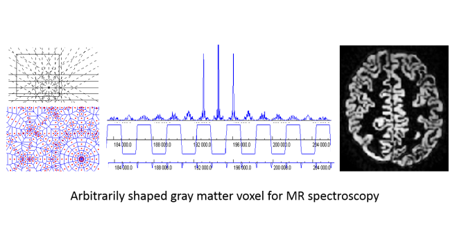
For MR spectroscopy, a technique to measure the concentrations of important metabolites, we have developed methods to acquire arbitrarily shaped target regions that can match anatomically or clinically defined target regions with minimal partial volume effects and, thus, enhance the impact and significance of the experiments. Other research projects deal with MR imaging with blood-oxygenation-level dependent contrast, the most important tool to investigate the function of the central nervous system in vivo, and aim to increase the temporal and spatial resolution and improve its applicability in the spinal cord, e.g. to investigate the processing of sensory input like pain.

In collaboration with research groups at the UKE and other universities, the developed and improved methods are applied in biomedical research and clinical studies in healthy volunteers and patients to evaluate their feasibility in practice and clinical setups. Finally, we provide methodological support for scientists performing MR experiments at the department's whole-body MR system, in particular for anatomical and functional measurements of the human brain.

  • Staff
  • Our current research projects are

    • the application of double-wave-vector or double-diffusion-encoded MR imaging to investigate tissue microstructure, e.g. to determine mean cell size or cell eccentricities

    • the development of improved MR imaging techniques for functional and diffusion-weighted imaging of the central nervous system, in particular for the investigation of the spinal cord

    • the measurement of anatomically defined target regions in MR spectroscopy to minimize partial volume effects, in particular for brain gray matter

    In these projects we apply

    • self-writeen pulse sequences for MR imaging and spectroscopy

    • numerical simulation of diffusion and the Bloch equations

    • model systems to test and validate the developed techniques

  • Lawrenz M, Brassen S, Finsterbusch J. Microscopic diffusion anisotropy in the human brain: age-related changes. Neuroimage 141, 313-325 (2016)

    Finsterbusch J, Sprenger C, Büchel C. Combined T2*-weighted measurements of the human brain and cervical spinal cord with a dynamic shim update. Neuroimage 79, 153-161 (2013)

    Finsterbusch J, Busch MG, Larson PE. Signal scaling improves the signal-to-noise ratio of measurements with segmented 2D-selective radiofrequency excitations. Magn. Reson. Med. 70, 1491-1499 (2013)

    Busch MA, Finsterbusch J. Eliminating Side Excitations in PROPELLER-Based 2D-Selective RF Excitations. Magn. Reson. Med. 68, 1383-1389 (2012)

    Finsterbusch J. Improving the performance of diffusion-weighted inner-field-of-view EPI based on 2D-selective RF excitations by tilting the excitation plane. J. Magn. Reson. Imaging 35, 984–992 (2012)

    Eippert F, Finsterbusch J, Bingel U, Büchel C. Direct Evidence for Spinal Cord Involvement in Placebo Analgesia. Science 326, 404 (2009)

  • Feroze Mohamed
    Thomas Jefferson University, Philadelphia, USA;
    Diffusion-weighted MR imaging of the pediatric spinal cord.

    Falk Eippert
    Max Planck Institute for Human Cognitive and Brain Sciences;
    Functional imaging of the spinal cord.

    Sune Jespersen
    University of Aarhus, Aarhus, Denmark;
    Non-Gaussian diffusion in the human brain.

    Peder Larsen
    University of California – San Francisco, San Francisco, USA;
    Spatially two-dimensional RF excitations.

    Julien Cohen-Adad and Julien Doyon
    Ecole Polytechniques, McGill University, Montreal, Canada;
    Diffusion-weighted and functional imaging of the spinal cord.

    Karl Maier
    University of Bonn, Bonn, Germany;
    Elastography of breast and brain tissue.

  • Our group has been supported by grants from the Deutsche Forschungsgemeinschaft and Wings for Life.